What are optic disc drusen?
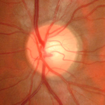
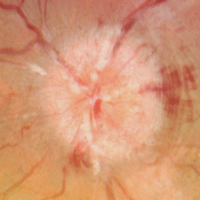
The optic nerve represents the cable connecting the eye to the brain, carrying all of the information from the retina that we need to see. As it exits the back of the eye, the optic nerve head normally appears as a yellow-pink circle with a discrete round edge. Certain serious conditions can cause the optic nerve to become swollen, including pseudotumor cerebri, optic neuritis, central retinal vein occlusion, infections, and brain tumors. Many of these conditions cause an increase in the pressure of the fluid surrounding the brain (cerebrospinal fluid). Bilateral optic nerve swelling from increased intracranial pressure is called papilledema.
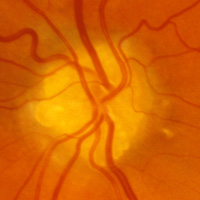
One of the more common conditions that can simulate papilledema is optic disc drusen. Drusen are an accumulation of calcium deposits within the nerve head, giving it a lumpy, irregular and elevated appearance. This is called pseudopapilledema. Drusen may be located near the nerve surface where they appear as yellow round or oval lumps or can be buried deeper in the nerve tissue where they can be very difficult to detect. Drusen generally become more visible with age. Optic disc drusen occasionally run in families as a dominant trait.

Normal optic nerve

Pseudopapilledema from optic nerve drusen

Swollen optic nerve from papilledema
What are the symptoms of optic disc drusen?
Most patients with optic disc drusen are totally asymptomatic and are diagnosed during a routine ocular examination. Patients may rarely notice decreased central vision or areas of peripheral vision loss.
How are optic disc drusen diagnosed?
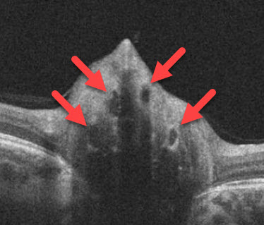
Superficial optic disc drusen are often easily found during a dilated eye exam. Buried disc drusen are more difficult to detect. Fundus autofluorescence, ultrasonography, and OCT scanning can be used to help identify buried drusen. However, it is sometimes extremely difficult to tell if a patient has pseudopapilledema from optic disc drusen or true papilledema from a serious medical problem. In these instances, MRI scanning and subsequent lumbar puncture may be necessary.

How are optic disc drusen treated?
Optic disc drusen are almost always asymptomatic and require no treatment. Patients need to be aware that their nerves are not truly swollen in case this is questioned on subsequent exams to avoid unnecessary invasive testing. Patients can rarely develop the growth of new blood vessels around the optic nerve (macular neovascularization as can develop in angioid streaks, age-related macular degeneration, choroidal rupture, degenerative myopia, idiopathic, and ocular histoplasmosis). Patients should therefore check their vision regularly with an Amsler grid and notify their eye doctor immediately if any changes occur.
View more retina images at Retina Rocks, the world’s largest online multimedia retina image library and bibliography repository.


