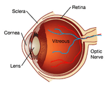
The human eye is like a camera where outside images are focused onto a piece of film. The cornea and crystalline lens are the lenses that focus the picture onto the eye’s film, the retina. The iris is the colored circle in the front of the eye. The black pupil, in the center of the iris, enlarges and contracts to regulate the amount of light entering the eye. The vitreous is a transparent gel filling the inside of the eye. The choroid is a system of blood vessels that covers the outer retinal surface, providing it with oxygen and nourishment. The sclera, or white of the eye, is a tough protective outer shell that corresponds to the body of a camera. The optic nerve carries the light images to the brain.

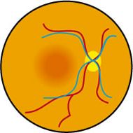
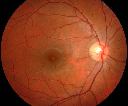
The macula is a small specialized area of the retina responsible for straight-ahead reading and driving vision. The retina reacts to light through a chemical process which then sends vision information through the optic nerve directly to the brain where the “picture” is processed.

Diagram of the normal retina and macula.

Photograph of the macular region.
Unlike a camera, the image obtained by the retina is not of uniform clarity or sharpness. Only the macula is sensitive enough to provide high-quality central vision. Any disease that affects the macula, such as diabetes, macular degeneration, macular hole, macular pucker, vitreomacular traction, ocular histoplasmosis, retinal detachment, or retinal vein occlusions can cause symptoms such as central blurriness or distortion.


